
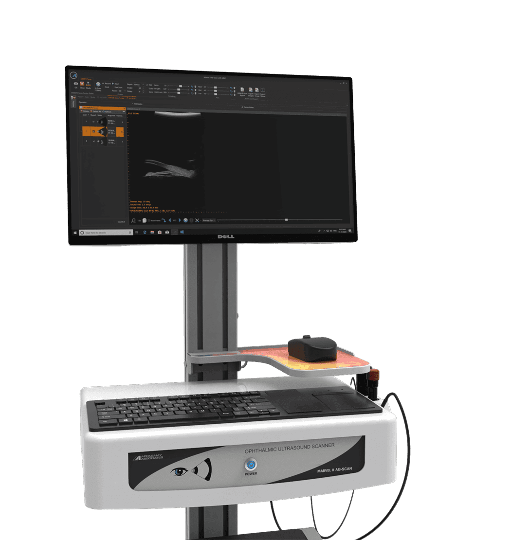
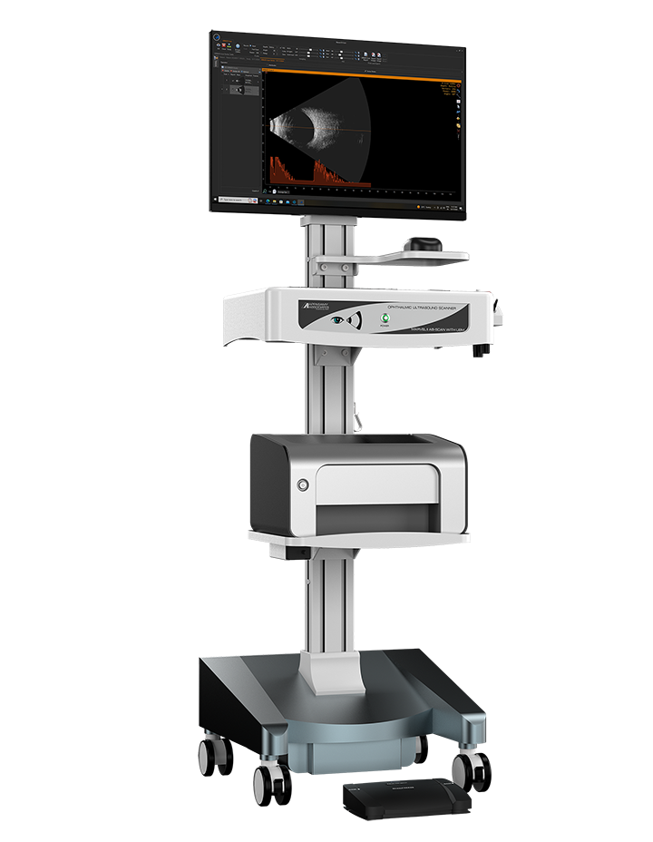


Marvel II AB – Scan With UBM
A SCAN


Enhanced Diagnosis
The MARVEL II AB scan with UBM uses the conventional A-scan pattern recognition program for the axial length measurement. It utilizes ultrasound waves of 10MHz frequency for the diagnosis. The built-in fixation LED for patient Target of Accuracy Reading.
Iol Calculator
The IOL calculator is easy to use and includes all formulas, including SRK – T, SRK – II, Holladay, Binkhorst, ( Hoffer Q : Price on Request ), Haigis, and Post Refraction Formulae. This system provides quick and accurate calculations.
B SCAN
High Resolution Imaging Technology
The MARVEL II AB scan with UBM produces reliable and completely focused B-mode image without any degradation of resolution. It has a unique technology which eliminates artifacts and allows deep penetration without any impact in image quality. It has a high resolution display system for effective diagnosis.


B-Scan Modes
There are 3 basic B-Scan modes provided by the equipment which enhances the diagnosis in the targeted area. The following are the types of modes for specific applications
Orbit Mode: Orbital muscle related Diagnosis can be done in this mode.
Vitreous Mode: Detailed Vitreous related Diagnosis can be done in this mode.
Retina Mode: In this mode the entire Posterior segment of the eye is viewed with greater details.

Good Image Quality
With the latest probe technology MARVEL II AB scan with UBM produces high resolution image for accurate diagnosis. There are 3 ranges of probes are available for B-scan
10 MHz: This probe allows deep penetration and provides low resolution image usually helps in viewing entire eye structure.
(.Or)
12 MHz: his probe allows deep penetration and provides low resolution image usually helps in viewing entire eye structure.
(.Or)
20 MHz: This probe produces high resolution image but penetration is shallow so this kind of probes are used in anterior view of posterior segment.
Advance Feature
Post Processing Gain: The specialized post processing gain setting allows the user to adjust the gain value even in the image freeze mode, whereas in other equipments this is possible only in live recording.
AB Scans Capability: The MARVEL II AB scan with UBM has an advance capability of displaying A vector as spike image for the corresponding B scan image recorded which simplifies the diagnosis.
Flood Fill Colour: The MARVEL II AB scan with UBM software is designed with unique feature which allows highlighting the grayscale image with a color for a specific segment to improve the efficiency of the diagnosis.
TGC Mode: In this setting, gain can be adjusted in a specific portion of the image Live (or) after capturing.
High Quality Image Recording
The MARVEL II AB scan with UBM allows recording 600 frames per cine(Total 600* 100 =6000 frames) by providing motion picture diagnosis.
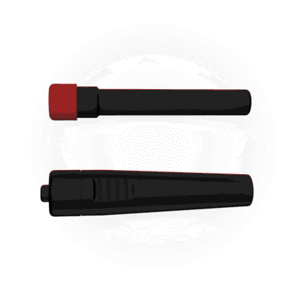
Sulcus To Sulcus Measurement
Sulcus to sulcus measuremnt is very crucial in determining the size of the IOL.Accurate sulcus to sulcus measurement can be obtained through proper probe placement.
Methods
The MARVEL II AB scan with UBM provides two different methods for anterior segment diagnosis.
Contact method: This is patient convenient method and it is useful for any infected cases.
Immersion method: This method is used regularly.


Measurement : Angle, Distance caliper, Area, Distance with A-Scan spikes, Probe orientation and Arrow tools.
Image Exporting: JPEG,BMP,TIFF,GIF,PNG
Report Exporting: PDF, HTML,MHT,RTF,
XLS,XLSX,CSV,
TXT and Image.
Data can Export to EMR software.
Laser Jet Printer.
Latest Micro form factor system with 2 TB HDD,8GB RAM & Windows 10 pro software

İlgili ürünler


A Scan Ophthalmic Ultrasound Scanner Model : Appascan Max
Fiyat & Teknik Bilgi İçin WhatsAppOther Features
-
Auto / Manual Gain
-
Automatic calculation of standard deviation and average axial length (series of 10 measurements)
-
Simultaneous display of 3 different IOL calculations
-
RTC shows the Date and Time throughout the life of the instrument.
-
Various user Programmable printouts Format
-
Measurement possible for Various Velocity of each segment.
-
*AME Formula - The new generation programmed software takes care of any Axial Length.


